joimax® Celebrates 20th Anniversary, Expands Education Program

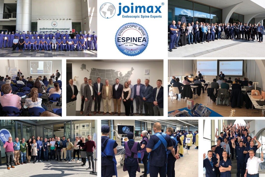
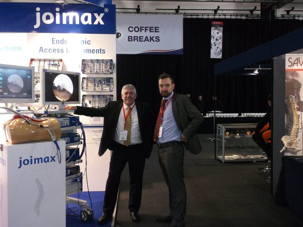
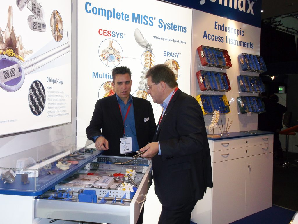
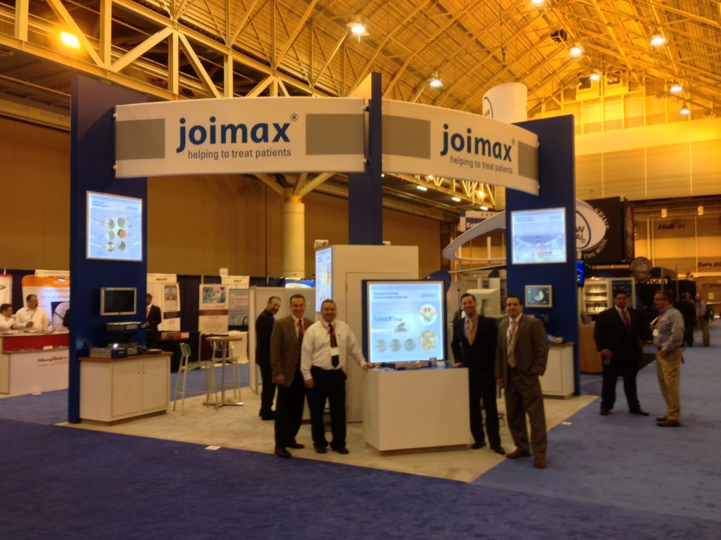
Karlsruhe (GER) — Founded 2001 in Germany, joimax®, the leading manufacturer of medical devices for endoscopic minimally invasive spine surgery, celebrates its 20th anniversary this year. In tandem with this milestone, the company expanded their educational program, ESPINEA®, furthering their global reach.

Despite COVID-19, ESPINEA® (Endoscopic Spine Academy) training programs, which are held in cooperation with the Royal College of Surgeons Edinburgh (RCSEd) and joimax®, were conducted all over the world in 2020, and continue to roll out in 2021. Due to travel restrictions, the programs were adapted to webinars, and remote support and training, totaling 60 sessions last year. By pivoting to online learning, approximately 3000 surgeons were reached in 2020, and 700 surgeons so far in 2021. ESPINEA® was founded by joimax® CEO and Founder Wolfgang Ries in 2019, and was accredited by the RCSEd last year. 

“Our goal is to be globally present online, as well as to provide direct local support,” says Ries. “Regardless of lockdowns in many countries, under the ESPINEA® platform, more than 120 surgeons were trained in onsite workshops this quarter, with 70 in the USA and Mexico, 35 in Europe, 15 in Asia, and many more through the joimax® CME-program partnership in China.

ESPINEA® Board Member Alastair Gibson, FRCSEd states: “These efforts demonstrate that joimax® and ESPINEA® can achieve anything together. I’m proud to be part of this impressive blend of online, on-site, and remote events, enabling a strong ESPINEA® community.”

To support the roll out, additional Coronavirus-compliant labs were integrated into the local, onsite education programs. In addition to the company’s existing labs in Karlsruhe and Irvine, California, more labs throughout the USA, including Houston, Minneapolis, New York City, Dallas, and Indianapolis, as well as the Surgical Skills Lab in Mexico City and ORSI Academy in Melle, Belgium, have been added.  Also, joimax® opened a branch office in Singapore; the Hong Kong office was opened in spring 2019.

“It is becoming increasingly important to have a personal, on-site presence in order to teach globally-valid learning content,” adds Ries. 

joimax launches the iLESSYS® Delta system for treatment of spinal stenosis at DGNC 2015 annual meeting

Karlsruhe, Germany – June 8, 2015 (BUSINESS WIRE) – The German company joimax®, developer of technologies and training methods for minimally invasive endoscopic spinal surgery, today announced it will introduce the company’s new iLESSYS® Delta system to the market at the 66th Annual Meeting of the German Society of Neurosurgery (DGNC) taking place in Karlsruhe, Germany from June 7-10.

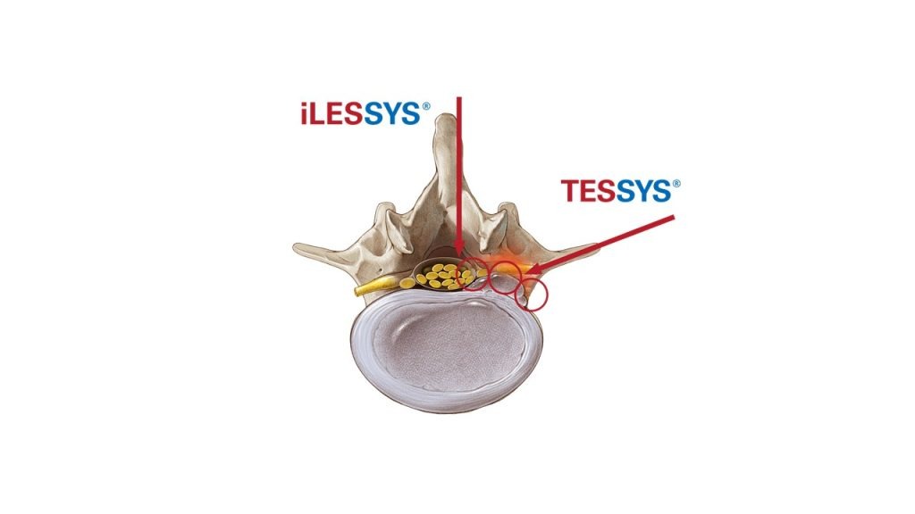
iLESSYS® Delta, an advanced development of the joimax iLESSYS® technology, is suitable for dorsal and dorso lateral treatment of central spinal canal stenosis. The iLESSYS® Delta system enables a large area to be decompressed by means of the interlaminar access, guided by endoscopic-assisted vision.

“Since the access to the spine is the same as with the already well-established iLESSYS® technique, usage is very straightforward.” said Dr. Guntram Krzok, orthopedic surgeon from Waltershausen, Germany after testing the new system at a joimax workshop early this year. “I was impressed by the new scope which delivers outstanding image quality. With the specialized instrument set, it is finally possible to achieve decompression of central spinal canal stenosis.”

The iLESSYS® Delta instrument set includes specially developed tools that enable extensive, yet gentle, tissue-sparing decompression. The 6 mm working channel of the new endoscope allows for the use of large shaver blades designed for bone resection and endo-kerrisons. An additional option is the plastic (PPSU) working tube for unimpaired X-ray examination.

“iLESSYS® Delta is a logical addition to our portfolio. Not only is the system suitable for endoscopic decompression, but it also allows the parallel implantation of cages. This way we can achieve dorsal decompression and stabilization in one step,” comments Wolfgang Ries, CEO and founder of joimax.

About joimax

Founded in Karlsruhe, Germany, in 2001, joimax® is one of the leading medical device companies in minimally invasive spinal surgery („joined minimal access“). The company’s U.S. subsidiary was established in Irvine, California, in 2005. The company is primarily focused on the development, production and marketing of technologies and methods for minimally invasive endoscopic spinal surgery. joimax is active in 40 countries around the globe and its methods have been successfully employed in approximately 130,000 surgeries. With a special focus on education, the company provides surgeons with specialized technique training through the three-step joimax CM3 education program. This program includes visitations, cadaver workshops and live-surgery support.

joimax® conducts an international Live Spinal Surgery Workshop in cooperation with the Medical University of Graz, Austria

Spine specialists take part at the joimax® endoscopy workshop with live spinal surgeries

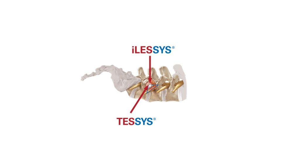
Karlsruhe, 14 March 2011: On 17 and 18 March 2011, the Medical University as well as the Institute for Anatomy in Graz, Austria, were the sites of a two-day international joimax® Workshop focused on the endoscopic resection of herniated discs utilizing the TESSYS® method. The first day, Dr. Wolfgang Kreil and Univ. Doz. Dr. med. Sandro Eustacchio of the Medical University Graz will operate on two patients with herniated discs using the TESSYS® method. The participating orthopaedic surgeons and neurosurgeons from Germany, Austria, Greece, Bulgary, Italy and Macedonia will experience the surgery procedure firsthand. While operating on the patients, one of the operating surgeons will explain each step of the TESSYS® surgery and answer questions of the workshop attendees. Afterwards Dr. Wolfgang Kreil and Dr. Sandro Eustacchio will lead scientific presentations and panel discussions on how the transforaminal endoscopic technique works and what their experiences have been, while discussing several case examples.

On the second day, joimax® hosts an intensive cadaver training course at the Institute for Anatomy Graz. The practical training and hands-on learning experience gained at cadaver workshops such as this one is an important first step in the three-step joimax® CM3 Education Program. In this comprehensive program, experienced surgeons and application specialists train each workshop member to use the TESSYS® Method. The subsequent steps of the Education Program are visitations, followed by live surgeries accompanied by joimax® Medical Trainers and application specialists.

joimax® starts the official sale of its EndoLIF® Delta and DoubleWedge cages. All three members of EndoLIF® family CE approved.

Karlsruhe, GER– July 15, 2019 –joimax®, the Germany-based market leader of technologies and training methods for full-endoscopic minimally-invasive spinal surgery, announces the official sales launch of its complete EndoLIF® family. EndoLIF® is an instrument set for endoscopic-assisted minimally invasive lumbar interbody fusion. As already announced and showcased at earlier events, the EndoLIF® product line consists of three members now: the well-known EndoLIF® O/On-Cage, the EndoLIF® Delta Cage, and the latest EndoLIF® Double Wedge Cage. The complete EndoLIF® product family is now CE-approved and fully available. Moreover, the EndoLIF® O/On-Cage is FDA-approved.

All EndoLIF® Implants are manufactured using a 3D printing process, which ensures an open diamond cell structure. The cages are safely implanted via the guide wire and are fillable with bone or bone substitute material. With their rough and porous surface, the implants guarantee optimal bone ingrowth, stability and finally fusion.

The EndoLIF® Cage family provides the right implant for every patient and is the perfect solution for endoscopic spinal fusion. With the gentle and atraumatic access via gradual tissue dilatation, muscles remain intact leaving important biomechanical structures in place after surgery.

Like with any endoscopic procedure compared to open surgery, all advantages for fusion are now given with the EndoLIF® program also. With one EndoLIF® Instrument Set all three cages of the EndoLIF® family can be implemented.

Dr. Florian Zentz, (Munich, GER) emphasizes “the extraordinarily high primary stability of the EndoLIF® DoubleWedge-Cage”. He continues enthusiastically: “After insertion the cage sits very firmly in the intervertebral disc space and adjusts the angulation up to 18 degrees, almost directly enabling an optimal lordosis correction.”

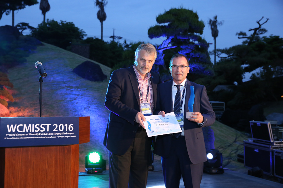
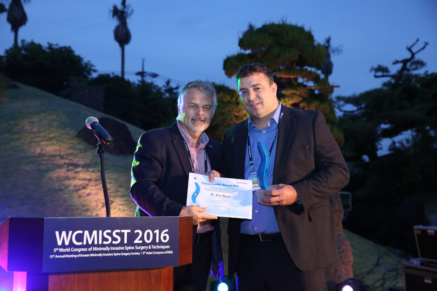
Dr. Ralf Wagner, (Frankfurt, GER) is also convinced of the obvious advantages: “The Delta Cage is ideal for the posterior lumbar interbody fusion (PLIF) offering a gentle posterior alternative, especially at level L5-S1 with its large interlaminar window.

A prime example of collaboration between China and Germany

Karlsruhe – joimax® opens the 3rd Training and Reference Center in Shanghai, China, at Changzheng Hospital, Tongji University, under the direction of Prof. Ye Xiaojian. The prestigious university and its affiliated Tung-Chee Hospital were founded in 1907 by the German government as its first major project in foreign cultural policy.

It is a prime example of Sino-German collaboration, which continues to this day at Tongji University and also at other centers in Chongqing at Xinqiao Hospital under the direction of Prof. Zhou Yue, and in Beijing at the General Hospital of the People’s Liberation Army (PLAGH) under the direction of Prof. Zhang Xifeng. The Training & Reference Centers provide targeted training to surgeons in endoscopic treatment methods on the spine as part of the joimax® Education Program. Above all, this features TESSYS®, the technique of transforaminal access to the lumbar spine for treatment of herniated discs and stenosis – an osseous narrowing of the spinal canal. Qualified and standardized training in these surgical techniques is essential. joimax® is therefore supporting the basic principle that patients should always receive high quality, reliable, and safe therapy in line with the very latest technology and scientific progress.

Zusammenarbeit_China_EN

Eurospine 2016: joimax® launches its new MultiZYTE® SI-Joint Endoscopic Therapy Set and its new Intracs® Intraoperative Navigation Tracking & Control System

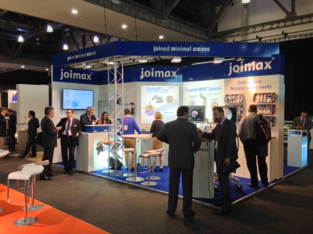
Karlsruhe – October 05, 2016 – joimax®, the global acting German developer and marketer of technologies and training methods for endoscopic minimally invasive spinal surgery, will again exhibit at Eurospine 2016 taking place from October 5 – 7 in Berlin. During the conference, joimax®will launch two new products – the MultiZYTE® SI Endoscopic Sacroiliac Joint Therapy Set and its new Intracs® Intraoperative Navigation Tracking & Control System.

MultiZYTE® SI Endoscopic Sacroiliac Joint Therapy is developed for the treatment of the Sacroiliac Joint Syndrome (SIJS), which has been rediscovered as a major back pain generator in recent years. This kind of pain is commonly felt in the lower back, the gluteal region, the hip and thigh and can even radiate into the leg and foot. Until the 1930s, SI-Joint was the main reason for low back pain, but since 1934 the focus has been on disc herniation. According to recent studies 15 to 25% of all low back pain is caused by the SI-Joint and up to 43% in patients having undergone lumbosacral fusion. MultiZYTE® SI joimax® now offers a well developed and lasting treatment option for this disease.

The Intracs® Intraoperative Navigation Tracking & Control System was developed in cooperation between joimax® GmbH Karlsruhe and fiagon GmbH in Berlin, both located in Germany. With the Intracs® system, joimax® instruments can be navigated directly at the tip using Fiagons “chip at the tip” technology which allows tracking of instruments with a diameter in the 1mm range. The Intracs ® Navigation and Monitor Unit is integrated in the joimax® Endoscopic Tower and all necessary tools are tailored for the TESSYS® (transforaminal) and iLESSYS® (interlaminar) procedures as well as for minimally invasive stabilization procedures, like EndoLIF® and Percusys®. The main features of the Intracs® system are needle navigation through Vector-Tip-Target processing tools, the merging of 2D X-Rays with 3D- images of a patient’s CT or MRI scans, and the constant orientation control of any endoscope used in joimax procedures.

“With these two new developments we are in the position to enhance our endoscopic minimally-invasive product portfolio, driving adoption rates to meet new heights. With the world unique Intracs® system, navigation is now faster, safer and more accurate than ever before using an electromagnetic (EM) field. Radiation exposure for both, patients and surgeons will be significantly reduced,” says Wolfgang Ries, CEO and founder of joimax®. “Additionally the learning curve for all spinal endoscopic procedures offered by joimax® will now be significantly reduced and the rapid expansion of all these techniques is now well layed-out,” he continues.

The Spine Movement – May 2012

Choll Kim, MD, PhD gives a breakdown of some of the most exciting presentations in MIS at ISASS12, and looks ahead to ISASS13 in Vancouver.

Minimally Invasive Surgery Was Huge at ISASS12

ISASS12 in Barcelona was a remarkable venue for numerous presentations related to minimally invasive spine surgery. Great interest both in the US and abroad continues to grow in this area of spine surgery: MIS made up the content for one-third of all the general sessions, with a sprinkling of individual presentations in other general sessions.

The range and complexity of the MIS topics proved broad. One of the most striking findings emerging in the literature is the marked decrease in infections when minimally invasive surgery is performed. Pfandistiener et al. from the Clinic for Spine Surgery with Scoliosis Center in Germany reported on infection rates between open and MIS posterior instrumentation. In 284 open surgeries, deep infections were seen in 2.46%. In contrast, 199 patients underwent percutaneous posterior instrumentation procedures without a single infection.

The use of minimally invasive strategies to treat ever more complex conditions were highlighted. Phillips and co-workers presented the prospective, multi-center results of 107 adult degenerative scoliosis with 24 month follow-up. Excellent clinical results and a low complications were encountered in this this challenging group of patients with advanced age and co-morbidities. Anand showed similar results in 33 patients from a single site.

The fusion rates of minimally invasive interbody fusion remains high. As shown in a meta-analysis by Gwinn et al. from Walter Reed Medical Center, the MIS TLIF technique enjoys a greater than 95% fusion rate.

Topics in endoscopic surgery continue to generate interest, as well–Nagahama et al. (Hokkaido University) and Schubert (Munich, Germany) showed continued success of this promising procedure in the hands of experienced endoscopic spinal surgeons.

Lingering questions regarding MIS were also addressed: Lee et al. (University of California, San Diego) quantitatively examined hip flexion strength after the popular transpsoas lateral lumbar interbody fusion (XLIF/DLIF/LLIF). As experienced by many surgeons, hip flexion strength decreased in most patients immediately postoperation, but returned to baseline within 2 weeks of surgery.

Cost effectiveness of open versus MIS TLIF was assessed by Phillips et al. on worker’s compensation patients; the cost of overall inpatient care was slightly less for MIS than for open.

Pimenta et al. advised appropriate caution when he described low fusion success rates of 2-level AxiaLIF procedures. In their review of 27 patients undergoing L4-S1 2-level AxiaLIF, only 22% of patients were considered to have solid fusion at 2 year follow-up.

Emerging MIS technologies were presented by a wide variety of groups. Regev et al. from Tel Aviv Sourasky Medical Center described the minimally invasive transforaminal, thoracic microscopic discectomy; Husain et al. from Loma Linda University described a percutaneous cervical transfacet fixation technique; Araghi et al. showed the results of the first 40 Guyer Lumbar Interbody Fusion (GLIF) procedures; MacMillan described a novel all transosseous approach to the L5-S1 disc, and both Julien (West Virginia University) and Sharma et al. (Manassas, VA) described the use of percutaneous stabilization for thoracolumbar fractures. It is heartening to see so many attempts to make the best of minimally invasive technology in this still pioneering area of spine surgery.

Santos et al. (University of Minnesota) showed that 3-D navigation may be used to improve safety and accuracy of percutaneous pedicle screw placement. Finally, the potential changes in healthcare delivery were highlighted by Smith and Christian (Las Vegas, NV) when they showed that selected MIS fusions may be performed in the ambulatory setting.

The diversity of topics was matched only by the excitement surrounding them. I’m looking forward to even more fascinating research by my colleagues at ISASS13 in Vancouver.

Choll Kim, MD, PhD
Spine Institute of San Diego

Virtual Global Symposium on Minimally Invasive Spinal Surgery Highlights Livestream Training Benefits

Karlsruhe, Germany — The Global Symposium of the ESPINEA® Academy in Karlsruhe, sponsored by joimax®, showcased how modern, advanced training for surgeons works. Earlier this month, over four days, more than 300 participants from around the world learned about the latest endoscopic surgical methods for spinal diseases. Via live demonstrations, twelve world-renowned spinal surgeons presented case studies, gave tips and tricks, and allowed people to look over their shoulders at endoscopic videos while operating on cadavers. The livestream made it possible for participating surgeons to understand exactly how endoscopic operations are carried out.

“You can only get closer if you do it yourself,” said one participant.

Endoscopic spinal surgery is developing into the gold standard for back problems that need surgery,” said Wolfgang Ries, the academy’s founder and chairman. “A large number of studies show it is superior to conventional methods of open spine surgery. Unfortunately, this technique is still not taught in most training centers around the world”.

ESPINEA® fills this gap with an extensive training program. Webinar formats are unavoidable in the Coronavirus period. With today’s livestream technologies, ESPINEA® was able to provide real-time, interactive surgical training, zooming in on how its surgical instruments can be used successfully.

“The participants’ enthusiasm is confirmation that we need to further develop online trainings,” said Ries. “While virtual events cannot replace face-to-face instruction — after all, it’s learning by doing — they provide an excellent alternative”.

ESPINEA® Academy was founded by joimax® in February 2019. A month later, joimax® CEO and Founder Wolfgang Ries held its first faculty and user meeting at the Paracelsus Medical University in Salzburg. Since then, ESPINEA® has been accredited by the oldest surgical college in the world, the Royal College of Surgeons Edinburgh. Participants at events such as the Global Symposium can earn medical training credits for Continuing Professional Development (CPD points). Points can be applied to medical license renewals.

Underscoring the importance of virtual learning, three ESPINEA® board members chaired the symposium: Menno Iprenburg from Iprenburg Spine Clinic in Veenhuizen, Netherlands; Sang-Ho Lee, president of Wooridul Spine Hospital, Korea; and J. N. Alistair Gibson, Royal College of Surgeons in Edinburgh. The participants concluded, “It was worth taking part just to hear these luminaries in person. But the best part was the opportunity to see the operation as if you were performing it yourself.”

Contact:
joimax® GmbH 
Nicole Read
info@espinea.org

joimax® faculty member Dr. Albert Telfeian performs 1st endoscopic removal of a thoracic spinal tumor

We are proud to share this story about our joimax® faculty member Dr. Albert Telfeian and how he was able to help 17-year old Jenelle “Nelly” Camara who was suffering from a spinal tumor.

Already in 2014, Dr. Telfeian removed a thumb-sized tumor pressing the girl’s spine cord. The pathology was sent for identification but even the best U.S. labs weren’t able to figure out what the tumor was. Camara underwent radiation therapy and was free of symptoms for a while. But the tumor resumed growth soon and she and her family returned to Dr. Telfeian at Hasbro Children’s Hospital in Providence.

This time the surgeon decided to use an endoscopic approach with the girl under local anaesthetic. With Camara being awake during surgery, Dr. Telfeian was able to ask her to move her legs which is an important control for the surgeon operating in such close proximity to the spinal cord. With a tiny 8-millimeter incision on her back and specialized endoscopic instruments he was able to do a biopsy on the tumor without removing bony material like in the first surgery.

Within a week the family received a diagnosis which now allows a targeted therapy for Camara who will be able to return to school and cheerleading in fall this year.
Read all the details about how endoscopy helped to identify Camara’s tumor in this article.

NBC also featured the story in NBC 10 News. 

A scientific paper (case report) about the procedure was published online in the journal, Clinical Neurology and Neurosurgery, and will appear in the July 2015 issue.

joimax® goes HD: HD Technology for superior safety in endoscopic spinal surgery

Camsource®HD camera systems, HD endoscopes and HD flat screen monitors by joimax® at the 5th German Spine Congress (5. Deutscher Wirbelsäulenkongress) in Bremen, Germany

Karlsruhe, December 2010: The 5th German Spine Congress (5. Jahrestagung der Deutschen Wirbelsäulengesellschaft (DWG) will take place from 16 to 18 December 2010 at the Congress Centre in Bremen. joimax® is proud to present the innovative HD 5-in-1 complete system at their booth no.20: The C-Camsource®HD Twister offers state-of-the-art HD technology and works with both the joimax® HD endoscopes and HD flat screens producing an extremely precise and sharp picture and video imaging for endoscopic spinal surgery.

This is convincing technology for spinal surgery experts like Dr. Michael Schubert, Apex-Spine Center, Munich, Germany or Dr. Ralf Wagner, Frankfurt, Germany: „High resolution like this allows very precise working and provides the necessary safety required when operating – especially in the field of endoscopic spinal surgery.“ With this integrated combination of HD camera technique, HD endoscopes and HD flat screen monitors joimax® is the first manufacturer to offer a completely tuned and expandable HD system of highest imaging quality and picture resolution.

Both surgeons value the C-Camsource® HD Twister as a 5-in-1 system with its integrated xenon light source and picture rotation function; “There is optimal lighting at all times and with the Twister, image details can be zoomed in and rotated”, “and thanks to the imaging function and the diaphragm and mask function, optimal orientation in the operation area is possible.”
The HD 5-in-1 complete system has also a text generator in order to document each step of the surgery and it offers direct and simple data storage on a USB data carrier. “It was important for us to offer the surgeon a perfectly coordinated as well as a safe complete system“, says Wolfgang Ries, CEO and founder of joimax®, “for the best possible treatment of patients – without any compromises.”

Advantages of the C-Camsource® HD-Twister complete system and the joimax® HD endoscopes

– Best possible resolution due to High-Definition-Technology
– First-class brightness, brilliance of the pictures due to optimal co-ordination of the endoscope,
camera and screen as tuned system
– Unique image rotation, zoom and picture movement function
– As much system integration as sensible, possible and necessary
– Functions for intuitive orientation, marking and documentation

Advantages of the HD flat screen monitors by joimax®

– HD flat screen monitors in either 24“ or 42“ with an angle of
178 degrees for an exceptionally good view and safe operating
– Brilliant images in high resolution: 1.920 x 1.080 pixels
– Picture in picture function; size and position adjustable
– Touch Control Panel, multi-language menu navigation