Eurospine 2016: joimax® launches its new MultiZYTE® SI-Joint Endoscopic Therapy Set and its new Intracs® Intraoperative Navigation Tracking & Control System

Karlsruhe – October 05, 2016 – joimax®, the global acting German developer and marketer of technologies and training methods for endoscopic minimally invasive spinal surgery, will again exhibit at Eurospine 2016 taking place from October 5 – 7 in Berlin. During the conference, joimax®will launch two new products – the MultiZYTE® SI Endoscopic Sacroiliac Joint Therapy Set and its new Intracs® Intraoperative Navigation Tracking & Control System.

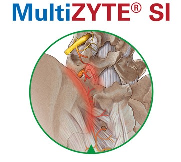
MultiZYTE® SI Endoscopic Sacroiliac Joint Therapy is developed for the treatment of the Sacroiliac Joint Syndrome (SIJS), which has been rediscovered as a major back pain generator in recent years. This kind of pain is commonly felt in the lower back, the gluteal region, the hip and thigh and can even radiate into the leg and foot. Until the 1930s, SI-Joint was the main reason for low back pain, but since 1934 the focus has been on disc herniation. According to recent studies 15 to 25% of all low back pain is caused by the SI-Joint and up to 43% in patients having undergone lumbosacral fusion. MultiZYTE® SI joimax® now offers a well developed and lasting treatment option for this disease.
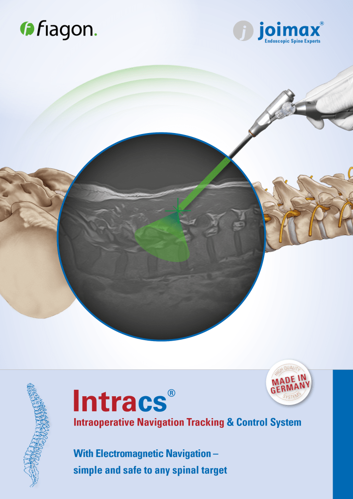
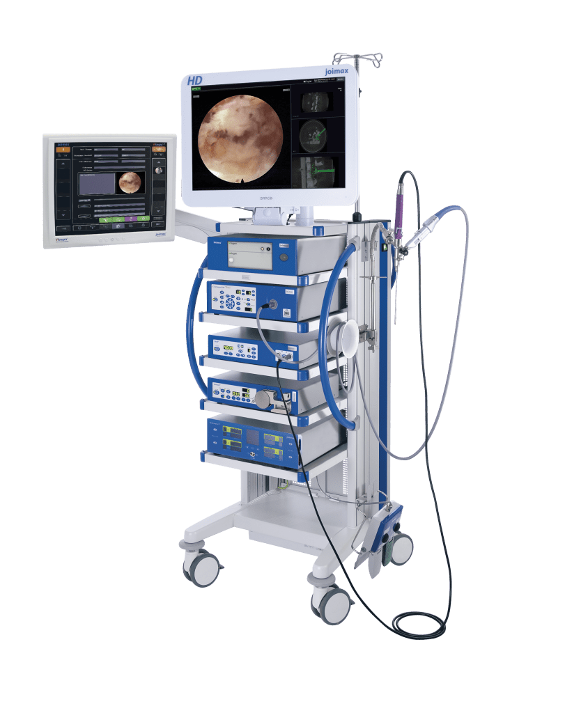
The Intracs® Intraoperative Navigation Tracking & Control System was developed in cooperation between joimax® GmbH Karlsruhe and fiagon GmbH in Berlin, both located in Germany. With the Intracs® system, joimax® instruments can be navigated directly at the tip using Fiagons “chip at the tip” technology which allows tracking of instruments with a diameter in the 1mm range. The Intracs ® Navigation and Monitor Unit is integrated in the joimax® Endoscopic Tower and all necessary tools are tailored for the TESSYS® (transforaminal) and iLESSYS® (interlaminar) procedures as well as for minimally invasive stabilization procedures, like EndoLIF® and Percusys®. The main features of the Intracs® system are needle navigation through Vector-Tip-Target processing tools, the merging of 2D X-Rays with 3D- images of a patient’s CT or MRI scans, and the constant orientation control of any endoscope used in joimax procedures.

“With these two new developments we are in the position to enhance our endoscopic minimally-invasive product portfolio, driving adoption rates to meet new heights. With the world unique Intracs® system, navigation is now faster, safer and more accurate than ever before using an electromagnetic (EM) field. Radiation exposure for both, patients and surgeons will be significantly reduced,” says Wolfgang Ries, CEO and founder of joimax®. “Additionally the learning curve for all spinal endoscopic procedures offered by joimax® will now be significantly reduced and the rapid expansion of all these techniques is now well layed-out,” he continues.

The Spine Movement – May 2012

Choll Kim, MD, PhD gives a breakdown of some of the most exciting presentations in MIS at ISASS12, and looks ahead to ISASS13 in Vancouver.

Minimally Invasive Surgery Was Huge at ISASS12

ISASS12 in Barcelona was a remarkable venue for numerous presentations related to minimally invasive spine surgery. Great interest both in the US and abroad continues to grow in this area of spine surgery: MIS made up the content for one-third of all the general sessions, with a sprinkling of individual presentations in other general sessions.

The range and complexity of the MIS topics proved broad. One of the most striking findings emerging in the literature is the marked decrease in infections when minimally invasive surgery is performed. Pfandistiener et al. from the Clinic for Spine Surgery with Scoliosis Center in Germany reported on infection rates between open and MIS posterior instrumentation. In 284 open surgeries, deep infections were seen in 2.46%. In contrast, 199 patients underwent percutaneous posterior instrumentation procedures without a single infection.

The use of minimally invasive strategies to treat ever more complex conditions were highlighted. Phillips and co-workers presented the prospective, multi-center results of 107 adult degenerative scoliosis with 24 month follow-up. Excellent clinical results and a low complications were encountered in this this challenging group of patients with advanced age and co-morbidities. Anand showed similar results in 33 patients from a single site.

The fusion rates of minimally invasive interbody fusion remains high. As shown in a meta-analysis by Gwinn et al. from Walter Reed Medical Center, the MIS TLIF technique enjoys a greater than 95% fusion rate.

Topics in endoscopic surgery continue to generate interest, as well–Nagahama et al. (Hokkaido University) and Schubert (Munich, Germany) showed continued success of this promising procedure in the hands of experienced endoscopic spinal surgeons.

Lingering questions regarding MIS were also addressed: Lee et al. (University of California, San Diego) quantitatively examined hip flexion strength after the popular transpsoas lateral lumbar interbody fusion (XLIF/DLIF/LLIF). As experienced by many surgeons, hip flexion strength decreased in most patients immediately postoperation, but returned to baseline within 2 weeks of surgery.

Cost effectiveness of open versus MIS TLIF was assessed by Phillips et al. on worker’s compensation patients; the cost of overall inpatient care was slightly less for MIS than for open.

Pimenta et al. advised appropriate caution when he described low fusion success rates of 2-level AxiaLIF procedures. In their review of 27 patients undergoing L4-S1 2-level AxiaLIF, only 22% of patients were considered to have solid fusion at 2 year follow-up.

Emerging MIS technologies were presented by a wide variety of groups. Regev et al. from Tel Aviv Sourasky Medical Center described the minimally invasive transforaminal, thoracic microscopic discectomy; Husain et al. from Loma Linda University described a percutaneous cervical transfacet fixation technique; Araghi et al. showed the results of the first 40 Guyer Lumbar Interbody Fusion (GLIF) procedures; MacMillan described a novel all transosseous approach to the L5-S1 disc, and both Julien (West Virginia University) and Sharma et al. (Manassas, VA) described the use of percutaneous stabilization for thoracolumbar fractures. It is heartening to see so many attempts to make the best of minimally invasive technology in this still pioneering area of spine surgery.

Santos et al. (University of Minnesota) showed that 3-D navigation may be used to improve safety and accuracy of percutaneous pedicle screw placement. Finally, the potential changes in healthcare delivery were highlighted by Smith and Christian (Las Vegas, NV) when they showed that selected MIS fusions may be performed in the ambulatory setting.

The diversity of topics was matched only by the excitement surrounding them. I’m looking forward to even more fascinating research by my colleagues at ISASS13 in Vancouver.

Choll Kim, MD, PhD
Spine Institute of San Diego